April 2023 Newsletter
Pancreatic [Imaging] Problems?
The pancreas is a retroperitoneal organ situated deep within the abdomen and not easily accessible by physical examination. Pancreatic pathologies have a variety of presentations, which make their diagnosis challenging to physicians.1 Imaging plays a critical role in the evaluation of pancreatic diseases and provides valuable information to clinicians, thereby dictating crucial management decisions. Technological advancements in multidetector computed tomography (MDCT) and magnetic resonance imaging (MRI), coupled with innovations in 3-dimensional (3D) imaging capabilities, have revolutionized the role of imaging in managing patients with pancreatic disorders. Imaging is most often performed to manage patients with pancreatitis and suspected pancreatic mass lesions.2
April Highlight: Imaging Techniques
This month, we’re highlighting how ChiRhoStim can aid in imaging of the pancreas.
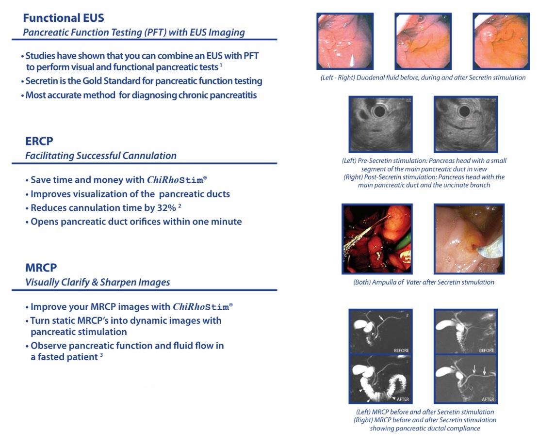
EUS
The advent of endoscopic ultrasound (EUS) provides ultra-high resolution images and exquisite details of pancreatic tumors, particularly in cystic lesions.
Additionally, EUS allows simultaneous tissue sampling of pancreatic lesions with EUS-guided fine needle aspiration (FNA). The acquisition and interpretation of these images are usually performed by interventional gastroenterologists.
EUS and endoscopic secretin pancreatic function test (ePFT) may be combined in a single endoscopic session (EUS/ePFT) to diagnose chronic pancreatitis (CP)3.
The two-sample ePFT serves as a useful adjunct to advanced endoscopic procedures such as endoscopic retrograde cholangiopancreatography (ERCP) and EUS. Patients with borderline or equivocal two-sample ePFT results should be considered for the 1-hour five-sample collection method.
ERCP
Endoscopic cholangio-pancreatography (ERCP) is a frequently employed second-step procedure to detect biliary and pancreatic abnormalities in patients with acute recurrent pancreatitis (ARP) of unknown aetiology. ERCP combines upper gastrointestinal (GI) endoscopy and x-rays to treat problems of the bile and pancreatic ducts. Doctors use ERCP to diagnose problems of the bile and pancreatic ducts if they expect to treat problems during the procedure, but prefer more noninvasive tests – tests that do not physically enter the body – which are safer and can also diagnose many problems of the bile and pancreatic ducts. For patients who undergo the procedure, a common post-ERCP complication is pancreatitis. Synthetic secretin is effective in reducing post-ERCP pancreatitis.4
MRCP
Magnetic resonance cholangiopancreatography (MRCP) has been used for non-invasive evaluation of the pancreas for several decades, providing excellent visualization of the pancreatic and biliary ductal systems. However, despite being the most accurate tool for pancreas imaging, the limiting nature of static imaging affects the providers’ assessment of pancreatic response to stimulation and real-time pancreatic function.
Secretin Enhanced magnetic resonance cholangiopancreatography (S-MRCP) allows physicians to assess the endocrine function of the pancreas in real time. The visualization of the pancreas is transformed from a static image (MRCP) to high temporal resolution images, which in turn is useful for getting a complete picture of the pancreatic response in physiologic condition and obtaining functional information.
Conference Update

17th Annual NPF Fellows Symposium | Hilton La Jolla Torrey Pines | La Jolla. CA
The NPF Fellows Symposium took place in La Jolla, CA this year and ChiRhoClin was so happy to have sponsored this years event. With a record number of attending fellows and phenomenal mentors, the symposium touched on topics including Pancreatitis, Pediatric Gastro, Oncology, and Surgery. Attending Fellows hailed from all across the United States and ChiRhoClin was honored to have been able to support the gathering of such exceptional individuals.
Future Events
May 5 – 7
Pancreas Club Annual Meeting | Palmer House Hilton Chicago | Chicago, IL
May 6 – 9
DDW Annual Meeting | McCormick Place Convention Center Chicago | Chicago, IL
Contact Wade Schoenecker for more information on where to find us!
References
1. Hrubran R, Wilentz R. The Pancreas. In: Kumar V, Abbas AK, Fausto N, eds. Robbins and cotran pathologic basis of disease 7th Ed. Philadelphia, PA: Elsevier Saunders; 2005:939-953.
2. “Imaging of the Pancreas: Part 1.” Imaging of the Pancreas: Part 1 • APPLIED RADIOLOGY, Applied Radiology, 4 Sept. 2013, https://appliedradiology.com/Articles/imaging-of-the-pancreas-part-1.
3. Stevens T, Dumot JA, Parsi MA, Zuccaro G, Vargo JJ. Combined endoscopic ultrasound and secretin endoscopic pancreatic function test in patients evaluated for chronic pancreatitis. Dig Dis Sci. 2010 Sep;55(9):2681-7. doi: 10.1007/s10620-009-1084-x. Epub 2010 Jan 26. PMID: 20101462.
4. Jowell PS, Branch MS, Fein SH, Purich ED, Kilaru R, Robuck G, d’Almada P, Baillie J. Intravenous synthetic secretin reduces the incidence of pancreatitis induced by endoscopic retrograde cholangiopancreatography. Pancreas. 2011 May;40(4):533-9. doi: 10.1097/MPA.0b013e3182152eb6. PMID: 21499206.