
Secretin Unlocks The Pancreas
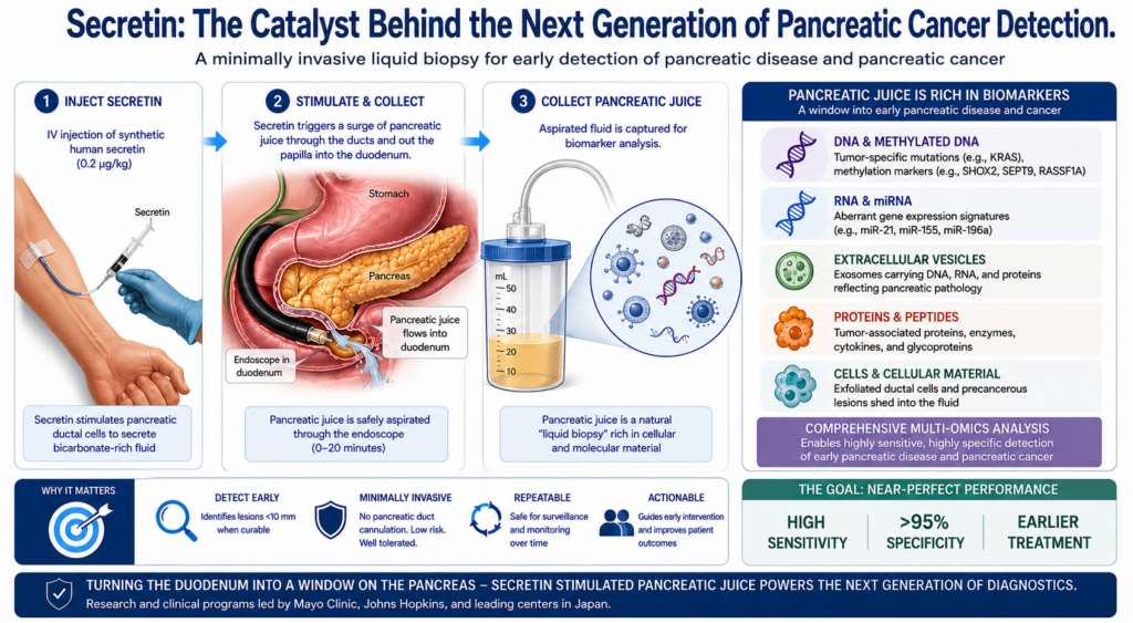
Secretin: The Catalyst Behind the Next Generation of Pancreatic Cancer Detection
How secretin-stimulated pancreatic juice is turning the duodenum into a window on early pancreatic disease — and why Mayo Clinic, Johns Hopkins, and Japan’s leading centers are building the future of diagnosis around it.
Pancreatic ductal adenocarcinoma (PDAC) remains one of the most lethal diagnoses in medicine, with five-year survival still below 13%. The reason is timing: most tumors are found only after they have spread. Yet when disease is caught early — lesions under 10 mm — five-year survival climbs toward 60%. The central problem has never been what to do about pancreatic cancer once found, but how to find it in time.
Conventional tools fall short of that goal. Serum CA 19-9 lacks the sensitivity and specificity to reliably flag resectable disease, and roughly 5–10% of patients carry a Lewis-antigen-negative phenotype that cannot produce the antigen at all. Cross-sectional imaging and even EUS/MRI surveillance routinely miss sub-centimeter cancers. What the field needs is a minimally invasive test that is highly sensitive with near-perfect specificity — and a growing body of evidence points to a familiar hormone as the key that unlocks it: secretin.
| Why Secretin — The Mechanism Secretin stimulates the pancreatic ductal epithelium to pour bicarbonate-rich fluid into the duct system and out through the papilla. That surge of pancreatic juice carries exfoliated ductal cells, DNA, RNA, extracellular vesicles and proteins directly into the duodenum — where it can be aspirated through a standard endoscope during EGD or EUS, without cannulating the pancreatic duct and without the pancreatitis risk of ERCP. In practice, synthetic human secretin is given as a 0.2 µg/kg IV dose and juice is collected within roughly 0–20 minutes. The result is a safe, repeatable “liquid biopsy” drawn from the organ’s own secretions. |
Mayo Clinic — Methylated DNA Markers Turn Juice Into a Liquid Biopsy
Mayo Clinic has anchored much of the modern case for secretin-stimulated collection. In a prospective, multicenter study, Engels, Majumder and colleagues assayed paired pancreatic juice and plasma from 88 biopsy-proven, treatment-naïve PDAC cases and 134 controls. A three-marker methylated-DNA panel (FER1L4, C13orf18, BMP3) recovered from secretin-stimulated, buffer-stabilized juice performed strongly on its own — and even better in combination with blood.
| 0.95 AUROC — juice panel + plasma CA 19-9 | 89% sensitivity, all PDAC stages (88% specificity) | 83% sensitivity, stage I/II disease |
The combined panel reached an AUROC of 0.95, significantly outperforming both the juice panel alone (0.87) and plasma CA 19-9 alone (0.91). At a fixed 88% specificity, sensitivity was 89% across all stages and 83% for early stage I/II disease — precisely the resectable window where detection changes outcomes. In a companion review in Gastroenterology (2025), Wallace, Majumder and coauthors frame secretin-stimulated duodenal collection as a platform that pairs methylated-DNA markers and extracellular-vesicle microRNAs with CA 19-9 to sharpen early detection and cyst surveillance. Parallel Mayo work has optimized juice collection and buffering to maximize tumor-derived DNA yield, and extended the same marker score to peritoneal-lavage staging (AUC 0.98), underscoring how far the methylated-DNA approach now reaches.
Johns Hopkins — Reading the Genetic Signal in Stimulated Juice
Johns Hopkins investigators (Goggins and colleagues) helped establish the founding principle behind the field: that secretin-stimulated pancreatic juice carries detectable, low-abundance driver mutations — KRAS, TP53, SMAD4 and GNAS — that mirror neoplastic change across the gland. Using highly sensitive sequencing, more than 1% mutant DNA has been found in the juice of roughly half of PDAC patients versus a small minority of those without cancer, and high-risk individuals can show an elevated mutational burden before any lesion is visible on imaging — a powerful signal for surveillance of familial and germline-risk patients.
That translational momentum now feeds the Pancreatic Cancer Detection Consortium (PCDC). In its 2026 blinded biomarker “bakeoff” (Clinical Cancer Research), the Johns Hopkins CA 19-9/FUT2/3 tumor-marker gene test was the single best-performing panel, reaching an AUC of 96.3 and significantly improving on CA 19-9 alone. The consortium’s design — head-to-head validation across Hopkins, Mayo and other centers — is exactly the rigor needed to move secretin-enabled and blood-based markers toward clinical use.
Japan — The Largest Prospective Validation to Date
Japanese researchers have delivered some of the most compelling clinical validation. In a seven-institution prospective study led from Osaka University and the National Cancer Center (Yachida et al., Annals of Surgery, 2025), secretin was given before EGD to stimulate pancreatic juice into the duodenum, where lavage fluid was collected with a dedicated catheter from 75 controls and 89 patients with resectable PDAC. A high-sensitivity assay for KRAS mutations then separated cancer from health with striking accuracy.
| Yachida et al. — Key Results ▪ AUC 0.934 (95% CI 0.904–0.964) for distinguishing resectable PDAC from healthy controls. ▪ 83.1% sensitivity (95% CI 71.7–91.2%) with specificity fixed at a demanding 100%. ▪ Significantly higher sensitivity than serum CEA and CA 19-9 (P < 0.0001) — and unaffected by the Lewis-negative CA 19-9 blind spot. |
Crucially, the test was validated in resectable-stage disease — the operable window that most influences survival. Complementary work at Tottori University (Takeda et al., Diagnostics 2022 and 2023) shows secretin’s reach beyond molecular markers: synthetic secretin-loaded pancreatic juice cytology raised cytologic sensitivity for malignant IPMN from 50.0% to 70.8%, and in a later series delivered 75% sensitivity, 100% specificity and 92.3% accuracy for PDAC. Secretin, in other words, strengthens both the genomic and the cytologic read.
An Expanding Secretin Toolkit
The same secretin-stimulated secretions are now feeding a widening menu of diagnostic strategies:
▪ Extracellular-vesicle microRNAs. Nesteruk et al. (Erasmus MC) profiled EV-miRNAs from secretin-stimulated juice; a panel of EV-miR-21/-25/-16 with serum CA 19-9 detected PDAC with an AUC of 0.91.
▪ Secretin-enhanced molecular imaging. Investigators at Massachusetts General Hospital used secretagogue-stimulated zinc-secretion MRI to detect PDAC and even track response to KRAS-G12D inhibition in preclinical models — probing the whole gland rather than hunting a tiny mass.
▪ Chronic pancreatitis & exocrine function. Secretin-stimulated EUS and endoscopic pancreatic function testing (DeWitt et al., Indiana/Dartmouth) refine the diagnosis of minimal-change chronic pancreatitis and exocrine insufficiency.
| The Bottom Line for Clinicians ▪ Secretin makes the duodenum an accessible sampling site — molecular and cytologic material without ductal cannulation or ERCP-level risk. ▪ Juice markers plus blood CA 19-9 consistently beat CA 19-9 alone — AUROC 0.95 at Mayo, AUC 0.93 in Japan, AUC 0.96 in the PCDC bakeoff. ▪ It flags the actionable window — resectable/early-stage cancer and high-grade dysplasia, sometimes before imaging shows a lesion. ▪ Convergent validation across three continents signals a maturing field poised to reshape surveillance of high-risk patients. |
| ABOUT SECRETIN IN THIS RESEARCH The studies discussed here rely on synthetic human secretin to stimulate pancreatic secretion. ChiRhoStim® (synthetic human secretin) is the formulation used in U.S. pancreatic function testing and referenced across this diagnostic literature, administered as a 0.2 µg/kg intravenous dose (see ChiRhoStim Package insert Updated-Long-Form-PI.pdf ). |
Selected References
1. Engels MML, Berger CK, Mahoney DW, et al. (incl. Majumder S). Multimodal Pancreatic Cancer Detection Using Methylated DNA Biomarkers in Pancreatic Juice and Plasma CA 19-9: A Prospective Multicenter Study. Clin Gastroenterol Hepatol. 2025;23:766–775.
2. Wallace MB, Majumder S, Storz P, van Hooft JE. Emerging Diagnostic Indications for Endoscopic Ultrasound. Gastroenterology. 2025 (in press).
3. Mills K, Taylor W, Berger CK, et al. (incl. Kisiel JB, Majumder S). Optimization of Endoscopically Collected Pancreatic Juice for Enhanced DNA Yield for Detection of PDAC. Mayo Clinic, Rochester MN (research presentation).
4. Mills K, Berger CK, Taylor WR, et al. (incl. Majumder S). Enhanced Molecular Staging of Pancreatic Cancer Using Methylated DNA Markers in Peritoneal Lavage Fluid. J Surg Oncol. 2026;134:257–262.
5. Oberg AL, Bamlet WR, Izmirlian G, et al.; Goggins MG; Pancreatic Cancer Detection Consortium. Pancreatic Cancer Detection Consortium Biomarker Bakeoff: A Phase II Blinded Biomarker Validation and Panel Discovery Study. Clin Cancer Res. 2026;32:2438–2448.
6. Yachida S, Yoshinaga S, Shiba S, et al.; Matsumoto K. KRAS Mutations in Duodenal Lavage Fluid After Secretin Stimulation for Detection of Pancreatic Cancer. Ann Surg. 2025 (publish ahead of print). DOI:10.1097/SLA.0000000000006645.
7. Takeda Y, Matsumoto K, Onoyama T, et al.; Isomoto H. Efficacy and Safety of Pancreatic Juice Cytology with Synthetic Secretin in Diagnosing Malignant Intraductal Papillary Mucinous Neoplasms of the Pancreas. Diagnostics. 2022;12:744.
8. Takeda Y, Matsumoto K, Onoyama T, et al. Pancreatic Juice Cytology Evaluations Using Synthetic Secretin and Serial Pancreatic Juice Aspiration Cytological Examination for the Diagnosis of PDAC. Diagnostics. 2023;13:1536.
9. Siddappa PK, Kong N, Lee A, Jiang Y, Park WG. From bench to bedside: pancreatic juice as a platform for biomarker discovery in pancreatic disease. Korean J Intern Med. 2026;41:15–30.
10. Nesteruk K, Levink IJM, de Vries E, et al.; Bruno MJ. Extracellular vesicle-derived microRNAs in pancreatic juice as biomarkers for detection of pancreatic ductal adenocarcinoma. Pancreatology. 2022;22:626–635.
11. DeWitt JM, Al-Haddad MA, Easler JJ, Sherman S, Slaven J, Gardner TB. EUS pancreatic function testing and dynamic pancreatic duct evaluation for the diagnosis of exocrine pancreatic insufficiency and chronic pancreatitis. Gastrointest Endosc. 2021;93:444–453.
12. Clavijo Jordan V, Sojoodi M, Moloudi F, et al.; Caravan P. Molecular Magnetic Resonance Imaging of Dysregulated Zinc Secretion Detects Pancreatic Ductal Adenocarcinoma Lesions and Response to KRAS-G12D Inhibitor Treatment. Cancer Res. 2025.
13. Ideno N, Mori Y, Nakamura M, Ohtsuka T. Early Detection of Pancreatic Cancer: Role of Biomarkers in Pancreatic Fluid Samples. Diagnostics. 2020;10:1056.
*This newsletter is an educational summary prepared for healthcare professionals in gastroenterology and pancreatology. It synthesizes findings from the peer-reviewed literature cited above and is not intended as medical advice, a treatment recommendation, or promotional labeling for any product. Diagnostic performance figures reflect the specific study populations and methods described in each source. ChiRhoStim® is a registered trademark of ChiRhoClin, Inc.
ChiRhoClin, Inc. · Clinical Update · Secretin & Pancreatic Diagnostics · 2026